01TAVR
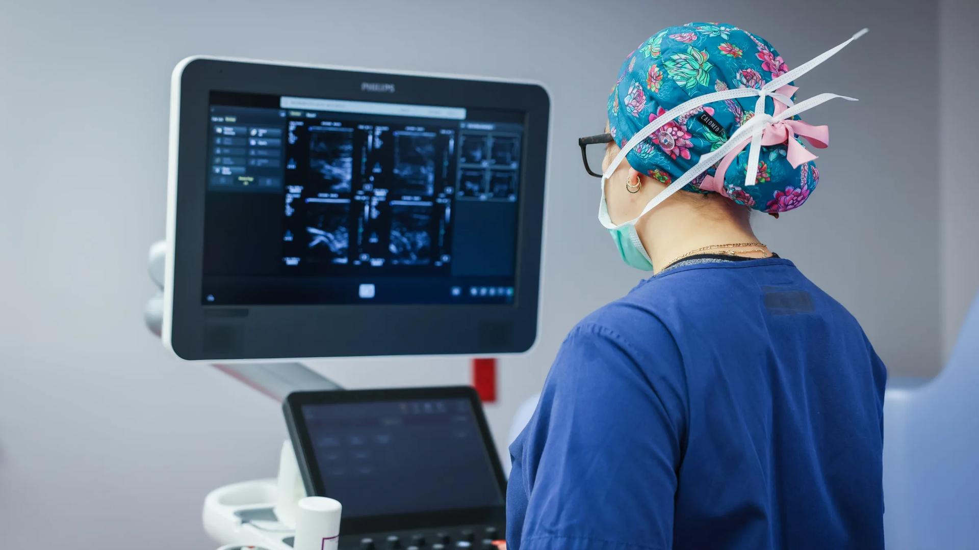
Here's the TAVR challenge. Your valve needs to deploy precisely in heavily calcified tissue and seal completely around irregular annuli across every variation you'll see clinically. We test valve expansion using fluoroscopy and echo to capture positioning stability and measure hemodynamic performance including leak rates and pressure gradients. We combine ISO 5840 testing with pulse duplicator measurements to address PMA and 510(k) pathway requirements.
02Valve Edge-to-Edge Repair (TEER)
Your clip grasps valve leaflets that must heal around the device. We evaluate tissue response at the grasp points, assessing inflammation, scarring patterns, and whether tissue remains viable without necrosis. Our pathologists examine leaflet healing quality using standardized scoring that FDA reviewers expect for chronic valve implants. This demonstrates your clip doesn't trigger excessive fibrosis or tissue damage that could compromise valve function over time.
03Mitral Valve Replacement
Your mitral valve anchors in soft tissue without calcification for support. We evaluate how atrial and ventricular tissue responds to anchoring forces, measuring inflammation, tissue compression, and healing at contact points. Our pathologists assess endothelialization on valve surfaces and tissue integration at anchor sites using standardized histological scoring. This shows FDA reviewers your anchoring mechanism achieves stable fixation without causing chronic inflammation or tissue necrosis.
04Tricuspid Valve Replacement
Your valve anchors in severely stretched right heart tissue that heals differently than left-sided structures. We evaluate tissue response at anchor points in dilated annuli, assessing inflammation, fibrosis, and whether pacemaker leads remain embedded safely. Our pathologists score endothelialization and tissue integration using protocols adapted for low pressure venous conditions. This demonstrates your device achieves stable fixation without damaging fragile atrial tissue or interfering with critical electrical leads.
05LAA Closure
Left atrial appendages vary wildly in size and shape. Your occluder has to seal across this variation without forming thrombus on the device itself. We test deployment precision in anatomically accurate LAA models and assess sealing effectiveness using flow visualization. We evaluate endothelialization progression over device surfaces. Does your device prevent strokes without causing them?